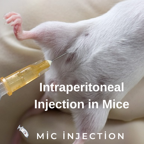
Intraperitoneal injection in mice can be performed in a number of ways. The head of the mouse should be held down to keep the organs from getting in the way of the needle. The needle should be inserted at a slight angle with the abdominal wall, and should be injected rapidly. Before completing the injection, it is important to check that there is sufficient fluid present in the peritoneal cavity.
Pain and distress measured after intraperitoneal injection
Pain and distress can be measured in laboratory animals, such as mice, after intraperitoneal administration of barbiturates, such as sodium pentobarbital. The AVMA Guidelines for the Euthanasia of Animals state that euthanizing animals by intraperitoneal injection of barbiturates is a safe and acceptable practice. In this study, pain and distress were assessed after intraperitoneal injection of sodium pentobarbital at concentrations of 5, 50, and 390 mg/mL. The animals were assessed for pain and distress by writhing, loss of the righting reflex, and time to death.
Mice were anesthetized by intraperitoneal sodium pentobarbital or saline. All mice were cervical dislocated, but four were excluded from the study after gross examination indicated that the injection had entered an organ. After the procedure, tissue samples were obtained and stained with hematoxylin and eosin. The tissues were then examined by a board-certified veterinary pathologist.
Pain and distress after intraperitoneal injection in mice is a sensitive measure of an animal’s response to an unpleasant stimulus. Besides the pain-like behavior, pain-related distress is often associated with increased grooming of the affected area or increased vocalizations. The distinction between pain and nociception highlights the critical difference in terminology. The treatment of animal pain must be based on the underlying causes of pain and distress.
Radioactivity distribution profile after intraperitoneal injection
The distribution of radioactivity in the mouse body is determined by using SPECT/CT images. Upon IP injection, peritoneal cavity radioactivity dominates the images within 1.5 hours. After this time, radioactivity in the bladder becomes visible and increases with time. The radioactivity level in other organs is minimal compared to the cavity. As a result, signals from other organs are overwhelmed. After 30 minutes, only the kidneys and bladder are visible.
In the present study, we measured the radioactivity distribution profile of 111In-Au@HSANP intraperitoneally administered to mice. During the first 2 minutes after injection, blood volumes were 0.6 mL higher than the injection volume. This hypertonicity was due to the presence of monocations in the labeling buffer at a concentration of 0.27 M, 1.7 times higher than that of isotonic saline. The remaining radioactivity in the mice’s body was detected at two to four hours after intraperitoneal injection.
AUC (area under the curve) of radioactivity after intraperitoneal injection in mice is a good predictor of bioavailability. The AUC (area under the curve) was 96 percent for the pretargeted group of mice, which translates to 96 percent bioavailability. As a result, the measured AUC after IP and IV injections should be similar.
Radioactivity distribution profile after intravenous injection
We have performed experiments in mice to assess biodistribution of radioactivity. We used a small animal SPECT/CT camera to visualize the distribution of radioactivity throughout the mouse body. In a single study, we injected mice with cMORF at different doses and determined the biodistribution profiles in the liver, lungs, and intestines. Afterward, we measured the remaining radioactivity in the entire body using a dose calibrator.
AUC was calculated for both routes of administration. We found that the AUC (area under the curve) for pretargeted radioactivity was similar (96%) when compared to AUC for the IP injection in normal mice. After intraperitoneal injection, we observed similar tumor accumulations and no change in the blood concentration time profile.
Our results showed that the kidneys were a key excretory organ for 111In-Au@HSANP. In the iv-injected group, the AUCs of the kidneys were 1.7-fold higher than those of the ip-injected group. The difference between the two groups was largely due to the a lower absorption in the peritoneum. The AUC of 111In-Au@HSANPS in mice after intraperitoneal injection was 3.55 +/1.00%ID/g, whereas AUC in the ip group was 0.41 +/0.06%.
111In-Au@HSANP retention in peritoneal cavity after intraperitoneal injection
To assess the efficacy of 111In-Au@HSANPS for cancer research, we performed an intraperitoneal injection of the compound into CT-26 mice with tumors. We found that 111In-Au@HSANS accumulated in the liver and spleen following ip administration. The clearance rate was 2.2 mL/h.
Au@HSANP was prepared by a slight modification from a previous study. The reaction mixture was centrifuged at 3000 g for 10 min to obtain a purified product. In addition, the radiochemical yield was >60%.
The authors found that 111In-Au@HSANPS retained approximately 90 percent of the administered dose. Their results suggest that the ip administration of Au@HSANPs has minimal cytotoxicity and may be a suitable drug delivery system for peritoneal carcinoma.
The average size of AuNPs was 22.5 + 5.9 nm and a polydispersity index of 0.16. The nanoparticles were also characterized by a high degree of opsonization, which occurs after uptake by macrophages and Kupffer cells. Their size and shape play a major role in determining their time in circulation, microdistribution in tumor regions, and retention. The NPs’ size and shape determine their ability to avoid first pass renal filtration. Those over 200 nm showed a slower clearance rate than the smaller particles.